Knee Pain and Exercise: Why Weak Glutes Are the Real Problem (And How to Fix It)
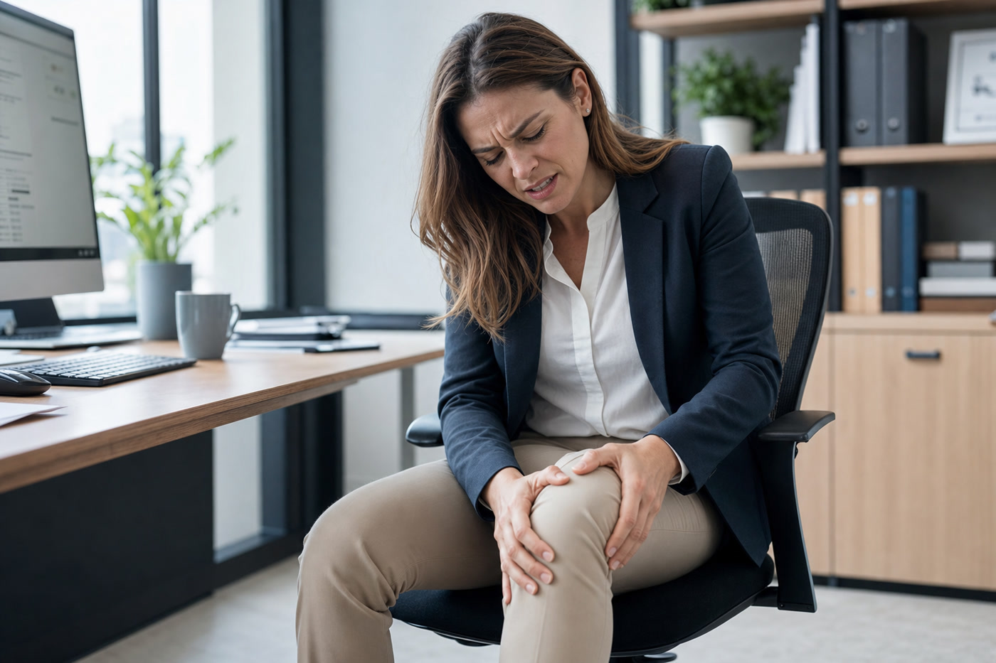
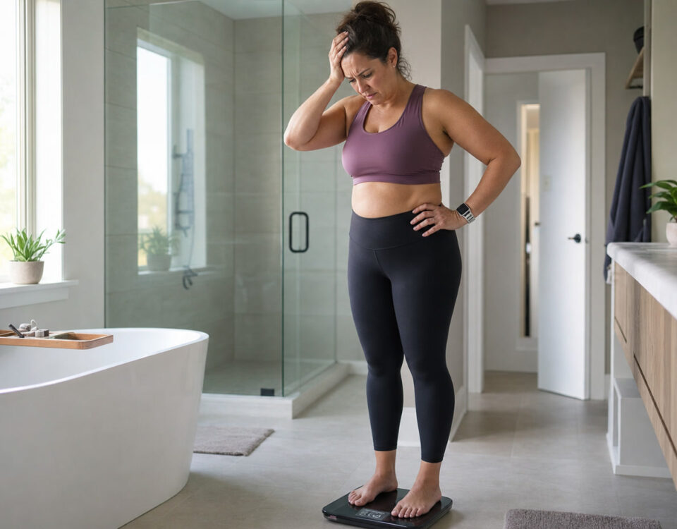
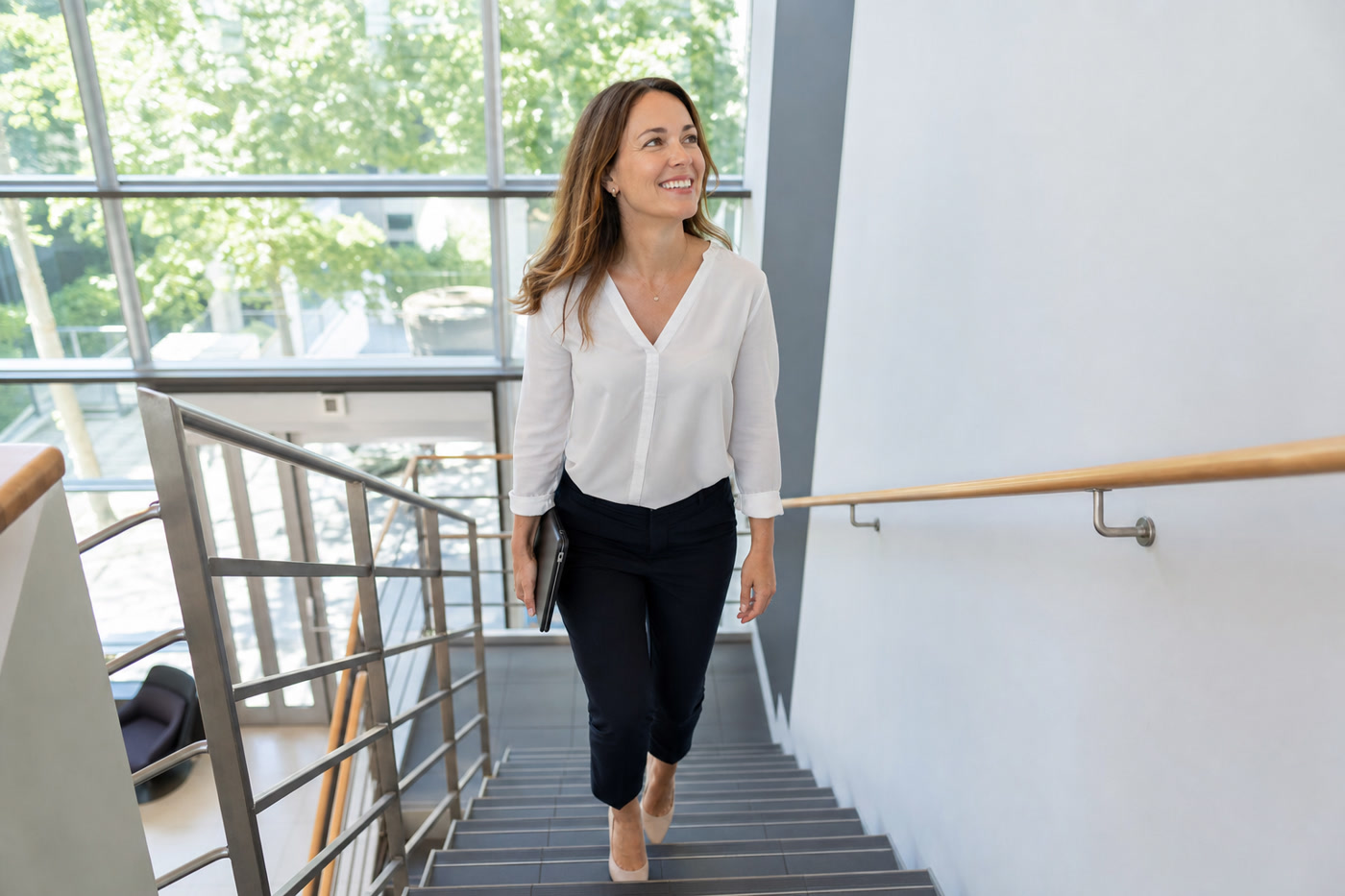
She has been dealing with knee pain for months. It started as a dull ache climbing the stairs from the SkyTrain. Now it greets her every time she stands up from her desk — that familiar throb behind the kneecap, the one that makes her think twice before every set of squats. She has seen her doctor. She has iced it. She has rested it. And she has been told, more than once, to “take it easy for a while.”
It is not getting better.
If this sounds familiar, here is what nobody has told you yet: the problem is almost certainly not your knee. The knee is where you feel it, but it is rarely where the dysfunction lives. For the majority of women experiencing anterior knee pain — particularly that nagging ache around or behind the kneecap — the real source is a chain of muscular imbalances that begins at the hip and, for some people, all the way down at the foot.
At TurnFit Personal Trainers in Vancouver, we work with hundreds of women navigating exactly this scenario — corporate professionals sitting 7+ hours a day, fitness beginners told exercise will hurt them, women who have been handed a rest prescription and a vague instruction to “strengthen their quads.” The science tells a very different story. Movement corrects this. The right movement, intelligently programmed, has been shown in randomised controlled trials to reduce patellofemoral knee pain by over 50% in six weeks.
This post explains exactly why this happens, who it affects (the statistics about women are striking), and what actually works — whether you train in our Kitsilano or Downtown Vancouver studios, or you are working with us online from anywhere in Canada.
The Scope of the Problem: Why Women, and How Many
Patellofemoral pain syndrome (PFPS) — the clinical term for the aching, grinding, or burning pain around the front of the knee — is one of the most common musculoskeletal complaints in the active population. But it is not evenly distributed. Women bear a disproportionate burden.
In a landmark epidemiological study drawing on over 2.1 million patellofemoral pain diagnoses across the United States, females accounted for 55% of all PFPS cases and 60% of chondromalacia patella diagnoses — the most direct form of cartilage-related patellofemoral pathology. This disparity was statistically significant (p < 0.001) across almost every age group, and was most pronounced in the 10–19 age bracket, where girls made up over 66% of cases. (Glaviano et al., 2015 — Int J Sports Phys Ther)
This is not a minor skew. It is a 2:3 ratio that demands explanation — and that explanation has profound implications for how we train, treat, and advise women with knee pain.
The Broader Knee Health Picture
PFPS is the acute, exercise-related end of the spectrum. Further along the continuum sits osteoarthritis (OA), the degenerative joint condition that is now Canada’s most prevalent chronic musculoskeletal disease:
| Group | Prevalence |
|---|---|
| Canadian females (20+) | 16.1% |
| Canadian males (20+) | 11.1% |
| Total Canadians with diagnosed OA | ~3.9 million |
Source: Public Health Agency of Canada, 2020
The knee is the most commonly affected joint in OA, and the pattern mirrors PFPS: women are more affected, at earlier ages, with greater functional impact. In British Columbia specifically, OA affects approximately 433,000 residents, and the knee accounts for the majority of cases requiring hip or knee replacement. (BC OA Survey, UBC Arthritis Research)
PFPS in your 30s and 40s is not inevitable. It is also not a life sentence. But the window to correct the underlying biomechanics is now — not after a decade of guarding, compensating, and deconditioning.
Why Are Women More Affected?
The sex disparity in PFPS is not random. Several anatomical and neuromuscular factors converge in ways that are more pronounced in women:
- Q angle: Women typically have a wider pelvis relative to femur length, creating a larger quadriceps angle (Q angle). This geometry places the kneecap under greater lateral stress and increases the tendency for patellar malalignment.
- Gluteus maximus inhibition during landing: Research indicates women demonstrate less gluteus maximus activation than men during landing tasks — a neuromuscular pattern that reduces the hip’s ability to control inward knee collapse. (Silvers-Granelli, 2021 — Int J Sports Phys Ther)
- Ligament laxity: Oestrogen influences ligament compliance, affecting joint stability across the hormonal cycle.
- ACL injury rates: Women have 3–8 times the ACL injury rate of similarly trained male athletes, and the same dynamic knee valgus pattern that drives PFPS also drives ACL tears. This is the same biomechanical problem on a severity spectrum. (Silvers-Granelli, 2021)
All of these factors are influenced by training. The gluteus maximus inhibition pattern is modifiable. The Q angle is fixed — but its consequence (dynamic knee valgus) is absolutely correctable through hip strengthening.
Ready to understand what’s actually driving your knee pain? TurnFit’s movement assessment identifies the exact links in your kinetic chain causing the problem.
The Real Cause — It Is Not Your Knee
Here is the biomechanics, explained plainly:
Your kneecap (patella) sits in a groove on the end of your femur called the trochlear groove. It is designed to glide straight up and down as you bend and extend your knee. When it glides correctly, pressure distributes evenly across the joint. When it tracks laterally — pulled off-centre by tight outer tissues or inward femoral rotation — the pressure concentrates on one area of cartilage. That is patellofemoral pain.
The question is: what pulls the patella off track? In most women with PFPS, the answer runs through the hip, not the knee itself.
The Gluteus Medius Inhibition Chain
The gluteus medius is the primary hip abductor — the muscle on the outer hip that keeps your pelvis level and prevents your knee from collapsing inward when you stand on one leg (which you do with every step you take). When the gluteus medius is inhibited, weak, or failing to fire at the right moment, a predictable cascade follows:
- Gluteus medius inhibition → the hip cannot control frontal-plane forces
- Femoral adduction and internal rotation → the thigh rotates inward under load
- Dynamic knee valgus → the knee collapses medially relative to the hip and foot
- Patellar maltracking → the kneecap is pulled laterally relative to the trochlear groove
- Patellofemoral pain → uneven cartilage loading, inflammation, anterior knee ache
This chain is well established in the literature. A 2025 study by Ho, Kissman, and colleagues published in Frontiers in Physiology found that gluteus medius central activation ratio (CAR) correlated significantly with Anterior Knee Pain Scale (AKPS) function scores in people with PFP (r = 0.584, p = 0.038) — meaning the more a person’s gluteus medius was actually firing, the better their knee function. Wide variability in gluteus medius activation (ranging from 84.4% to 98.3%) in PFP patients suggests a subgroup with meaningful inhibition whose symptoms would respond well to targeted re-activation work. (Ho et al., 2025 — Front Physiol)
The Gluteus Maximus Factor: ACL Risk and Knee Valgus
While the gluteus medius controls frontal-plane hip stability, the gluteus maximus controls hip extension and external rotation — and its inhibition has its own damaging consequences. Women demonstrate less gluteus maximus activation during landing tasks than men, contributing to the greater degree of dynamic knee valgus that predisposes female athletes to ACL injuries at 3–8 times the male rate. (Silvers-Granelli, 2021)
Research from the Journal of Experimental Orthopaedics confirms that gluteal muscle strength deficits directly influence dynamic knee valgus, and that solid glute strength is associated with substantially fewer knee valgus episodes during demanding tasks like jump landings — the same tasks that put the ACL under the greatest stress. (Losciale et al., 2022 — J Exp Orthop)
The bottom line: your glutes protect both your kneecap (PFPS) and your ACL (tear risk). Strengthening them is not optional — it is the cornerstone of knee health for women.
Hip Abduction Strength and Knee Valgus: What the Research Shows
A comprehensive scoping review of 29 studies examined the relationship between hip abduction strength and dynamic knee valgus. The consistent finding: stronger hip abductors produce measurably less knee valgus during squatting, landing, and walking — directly reducing patellofemoral joint stress. This is the mechanism by which hip strengthening resolves knee pain without ever touching the knee directly.
This is not a fringe finding. It is the basis of the current clinical practice guidelines for PFPS from the Journal of Orthopaedic and Sports Physical Therapy (Witvrouw et al., 2019 — JOSPT), which recommend hip strengthening as a core component of evidence-based PFPS management.
The Desk Worker Factor
If you work at a computer for most of the day — whether in a downtown Vancouver office tower or at a home desk in Point Grey — your working environment is actively working against your knees.
Here is why:
The Sitting–Glute Inhibition Loop
Prolonged sitting places the hip in a position of sustained flexion. When the hip flexors are held in a shortened position for hours, reciprocal inhibition reduces the neural drive to the opposing gluteal muscles. This is not a theory — it is a well-documented neuromuscular phenomenon. The result: a typical desk worker’s glutes are less neurologically “available” for immediate activation when they stand and begin moving.
This contributes directly to the gluteus medius inhibition chain described above. You get up from your chair, the glutes are slow to fire, the knee valgus pattern appears, and the patellofemoral joint takes the load it was not designed to bear.
Anterior Pelvic Tilt and the Knee Pain Connection
Prolonged sitting also encourages anterior pelvic tilt (APT) — the forward tilting of the pelvis that creates an exaggerated lumbar curve and places the hip and knee in suboptimal alignment for weight-bearing activity. This is not merely a postural cosmetic issue. A published study specifically examining the relationship between APT and PFPS found a statistically significant moderate positive correlation between anterior pelvic tilt angle and pain intensity in PFPS patients (r = 0.409, p = 0.009). In plain terms: the greater the forward tilt of the pelvis, the more intense the knee pain. (Al-Qattan et al. — J Biomed Arthritis Rehabil)
The Sitting Hours Data
Research on occupational sitting and knee pain has produced consistent findings:
- Office workers with knee pain sit an average of 6.72 hours per day — substantially above the threshold at which musculoskeletal risk increases
- Prolonged sitting has been shown to produce MSD symptoms in the thighs and knees of office workers, alongside more commonly discussed areas like the lower back and shoulders (Daneshmandi et al., 2017 — J Lifestyle Med)
- Approximately 3.9 million Canadians live with diagnosed osteoarthritis, the majority knee-related, and women are affected at 16.1% versus 11.1% for men — a gap that widens with age (Public Health Agency of Canada, 2020)
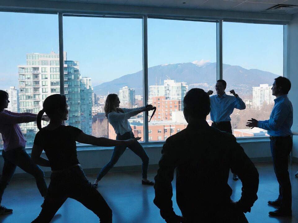
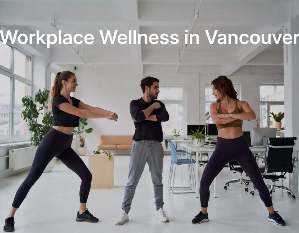
For TurnFit’s corporate wellness clients, this data is directly actionable. Desk workers are not suffering from bad luck; they are suffering from a preventable biomechanical environment that responds well to structured intervention. A corporate wellness programme that addresses hip mobility, glute activation, and movement patterning can meaningfully reduce workplace knee pain — and the productivity, absenteeism, and long-term health costs that come with it.
Does your team spend most of their day at a desk? TurnFit’s corporate wellness programmes address the specific musculoskeletal patterns that develop from desk work — including the knee pain that nobody talks about.
The Foot Foundation: The Kinetic Chain from the Ground Up
The gluteus medius gets much of the attention in PFPS research, and rightly so. But for some women, the chain of dysfunction does not begin at the hip — it begins at the foot.
Your foot is the first point of contact with the ground, and the forces it absorbs and transmits travel directly upward through the ankle, tibia, knee, and hip. When the foot over-pronates — the arch collapses and the heel rolls inward — the mechanical consequences ripple upward:
- Subtalar pronation → the heel bone rolls inward
- Tibial internal rotation → the shin bone rotates medially, coupled to subtalar motion
- Medial pull on the patellar tendon → disrupts kneecap tracking in the trochlear groove
- Patellofemoral malalignment → asymmetric joint loading and pain
This cascade has been studied extensively. Research by Barton and colleagues (2011) found that individuals with PFPS demonstrate earlier peak rearfoot eversion during gait compared to pain-free controls (p = 0.01), suggesting that more rapid foot pronation at heel strike is a measurable feature of patellofemoral pain patients — particularly women, who made up 81% of the PFPS group in that study. (Barton et al., 2011 — J Foot Ankle Res)
Studies examining foot posture and functional outcomes in PFPS consistently find that patients with greater foot over-pronation score significantly worse on the Kujala Anterior Knee Pain Scale — the gold-standard clinical measure of patellofemoral function — compared to those with neutral or supinated foot posture. The JOSPT 2019 Clinical Practice Guidelines now recommend foot orthoses for PFPS patients with greater-than-normal pronation, specifically combined with exercise therapy. (Witvrouw et al., 2019 — JOSPT)
Why Treating Foot Pronation Alone Is Not Enough
An orthotic addresses the foot end of the chain but does not strengthen the hip end. Similarly, hip strengthening alone may not fully eliminate a pronation-driven component. This is why TurnFit’s assessment evaluates the entire chain — foot mechanics, ankle mobility, tibial rotation, and hip strength — rather than focusing on a single joint. The right intervention targets the specific links in your chain that are failing.
For some clients, the primary driver is hip weakness; for others, it is foot mechanics; for many, it is both. A programme that does not assess all three levels will miss part of the problem.
What the Science Says About Fixing It
The research on hip strengthening for patellofemoral pain is extensive, consistent, and frankly, unusually decisive for musculoskeletal rehabilitation. Here is the evidence that matters most:
Ferber et al. (2015): The Multicenter RCT That Changed the Conversation
In the most rigorous trial to date on this question, Reed Ferber and colleagues at the University of Calgary led a multicenter randomised controlled trial with 199 participants (66% women, mean age 29) comparing a hip-and-core strengthening protocol (HIP group) to isolated knee strengthening (KNEE group) over 6 weeks. The results were clear:
- Both groups showed significant reductions in VAS pain scores and improvements in Anterior Knee Pain Scale function (p < 0.001 in both)
- The HIP group achieved pain resolution one week earlier than the KNEE group
- The HIP group showed significantly greater gains in hip abductor strength (p = 0.01) and posterior core endurance (p = 0.05)
- Baseline VAS scores in the hip group averaged 5.2/10; at 6 weeks, women in the hip group reduced to approximately 2.4/10 — a 54.7% reduction
(Ferber et al., 2015 — J Athl Train. PMID: 25365133)
Halabi et al. (2025): Meta-Analysis Confirms Hip + Knee Superiority
A 2025 systematic review and meta-analysis published in Musculoskeletal Care synthesised six RCTs involving 241 PFPS patients (96.3% female). The conclusion: combined hip and knee strengthening significantly outperforms knee strengthening alone for both pain reduction (SMD = -1.29, 95% CI [-1.98, -0.59], p = 0.0003) and functional activity (SMD = 0.99, 95% CI [0.22, 1.76], p = 0.01). These are large effect sizes, and the patient population was overwhelmingly female — making these findings directly applicable to the women reading this. (Halabi et al., 2025 — Musculoskelet Care. PMID: 39934098)
Supporting Evidence: Multiple RCTs and Systematic Reviews
| Study | Design | Key Finding |
|---|---|---|
| Ferber et al., 2015 | Multicenter RCT, n=199 | Hip protocol resolved pain 1 week earlier; ~55% VAS reduction in women at 6 weeks |
| Halabi et al., 2025 | Systematic review + meta-analysis, 6 RCTs, n=241 | Hip+knee strengthening significantly superior to knee-only for pain (SMD=-1.29) and function |
| Ho et al. (Kissman), 2025 | Case-control, gluteus medius CAR study | Higher gluteus medius activation = better knee function (r=0.584, p=0.038) |
| Barton et al., 2011 | Case-control, n=46 | PFPS patients show earlier rearfoot eversion (p=0.01) — foot pronation implicated |
| APT + PFPS correlation study | Correlational study | APT moderately correlated with PFPS pain intensity (r=0.409, p=0.009) |
| Witvrouw et al., 2019 | Clinical practice guidelines (JOSPT) | Hip strengthening + foot orthoses recommended as evidence-based PFPS management |
| Silvers-Granelli, 2021 | Review — ACL injury in women | Women have 3–8× greater ACL risk; gluteus maximus inhibition during landing key contributor |
| Glaviano et al., 2015 | Epidemiological, n=2,188,753 | Women = 55% of PFPS cases, 60% of chondromalacia cases (p<0.001) |
| Peña et al., 2024 | RCT, remote vs in-person | Online and in-person training produce equivalent strength and body composition outcomes |
What This Means in Practice
Six weeks of consistent, targeted hip strengthening — the kind that addresses your specific weak links in the kinetic chain — produces measurable, clinically significant improvements in knee pain. Not in a year. In six weeks. The evidence base for this is robust, replicated across multiple independent research groups, and consistently points to the hip as the key therapeutic target.
You do not have to wait for the pain to get worse. TurnFit’s online training programme delivers the same evidence-based hip strengthening protocols used in our Vancouver studios — from anywhere in Canada.
Why Most Approaches Fail
If the evidence is this clear, why do so many women with PFPS remain in pain for months or years? The answer lies in how knee pain is typically managed — and what most approaches get wrong.
“Rest It” Does Not Fix the Root Cause
Rest reduces inflammation and offloads the joint, which can temporarily ease symptoms. But rest does not address gluteus medius inhibition. It does not correct anterior pelvic tilt. It does not change the way you land, climb stairs, or squat. When you return to activity after a rest period, you return with the same biomechanical fault — and often with weaker hip and quad muscles than when you started, because they have not been used.
PFPS has a well-documented tendency toward chronicity when under-treated. Research tracking PFPS patients over 5–20 years found that 25–91% continued to report symptoms affecting daily life or physical activity — a range that largely reflects the quality of the rehabilitation undertaken. (Ferber et al., 2018 — J Athl Train)
Isolated Quad Strengthening Misses the Hip
The traditional prescription for PFPS — quad sets, straight-leg raises, terminal knee extensions — addresses only one side of the equation. These exercises can reduce pain and improve function, but the evidence now clearly shows that adding hip work produces superior and faster results. Treating the knee without the hip is like trying to fix a door hinge by only working on the door itself — when the frame is the problem.
Ice and Anti-Inflammatories Address the Signal, Not the Source
NSAIDs and icing manage the inflammatory response that PFPS produces, but they do not change the mechanical fault producing that response. They are appropriate short-term measures — but they are not a rehabilitation plan.
Generic Exercise Classes May Load the Knee Incorrectly
Group fitness classes and generic online workouts are not designed for your kinetic chain. If your gluteus medius is inhibited and you are performing high-volume squats, lunges, or step-ups without correction, you may be loading a dysfunctional pattern under progressive load — reinforcing the very mechanics causing your pain. This is why individualised assessment matters. The goal is not to avoid loading the knee; it is to load it correctly.
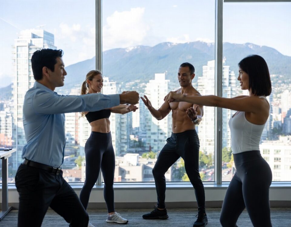
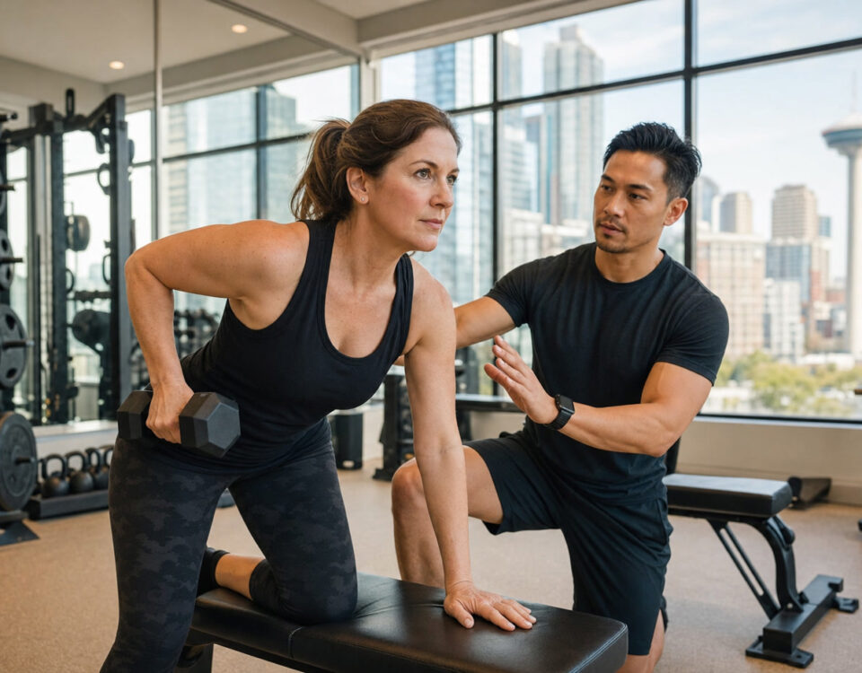
The TurnFit Approach: Assessment to Fix
TurnFit Personal Trainers has worked with hundreds of women in Vancouver experiencing exactly the knee pain described in this post. David Turnbull and the TurnFit team hold BCRPA certification, have earned 300+ five-star Google reviews, and have been awarded the Top Choice Award in Vancouver eight times — a track record built on producing real, lasting results.
The TurnFit approach to knee pain does not begin at the knee. It begins with a comprehensive movement assessment that maps your individual kinetic chain, then builds a programme targeting your specific weak points.
Step 1: Foot and Ankle Assessment
The assessment begins at the floor. TurnFit evaluates arch posture, subtalar pronation, and ankle dorsiflexion. Limited ankle dorsiflexion is a documented contributor to patellofemoral stress — when the ankle cannot dorsiflex adequately, the knee compensates by moving into greater flexion and valgus to accommodate movement. Foot over-pronation is assessed and, where clinically indicated, orthotic referral or specific foot intrinsic strengthening is incorporated into the programme.
Step 2: Pelvic Position and Hip Flexor Length
The assessment evaluates pelvic tilt and hip flexor length. Given the moderate positive correlation between anterior pelvic tilt and PFPS pain intensity (r = 0.409, p = 0.009), correcting APT through hip flexor lengthening and posterior chain strengthening directly addresses pain intensity — not just function. For desk workers, this step is almost always relevant.
Step 3: Gluteus Medius and Maximus Strength Testing
Hip abductor and extensor strength are assessed both quantitatively (manual muscle testing) and functionally (single-leg squat quality). This identifies the specific gluteal muscles requiring targeted work and establishes a baseline against which progress will be tracked.
Step 4: Dynamic Movement Screen
TurnFit observes you moving — squatting, stepping, landing, walking up and down stairs. This is where the biomechanical fault becomes visible: the knee caving inward, the trunk leaning, the pelvis dropping. These real-time observations confirm the assessment findings and identify the precise movement patterns to correct.
Step 5: Personalised Hip-Dominant Programme
From the assessment, TurnFit builds a progressive, individualised programme. The programme typically includes:
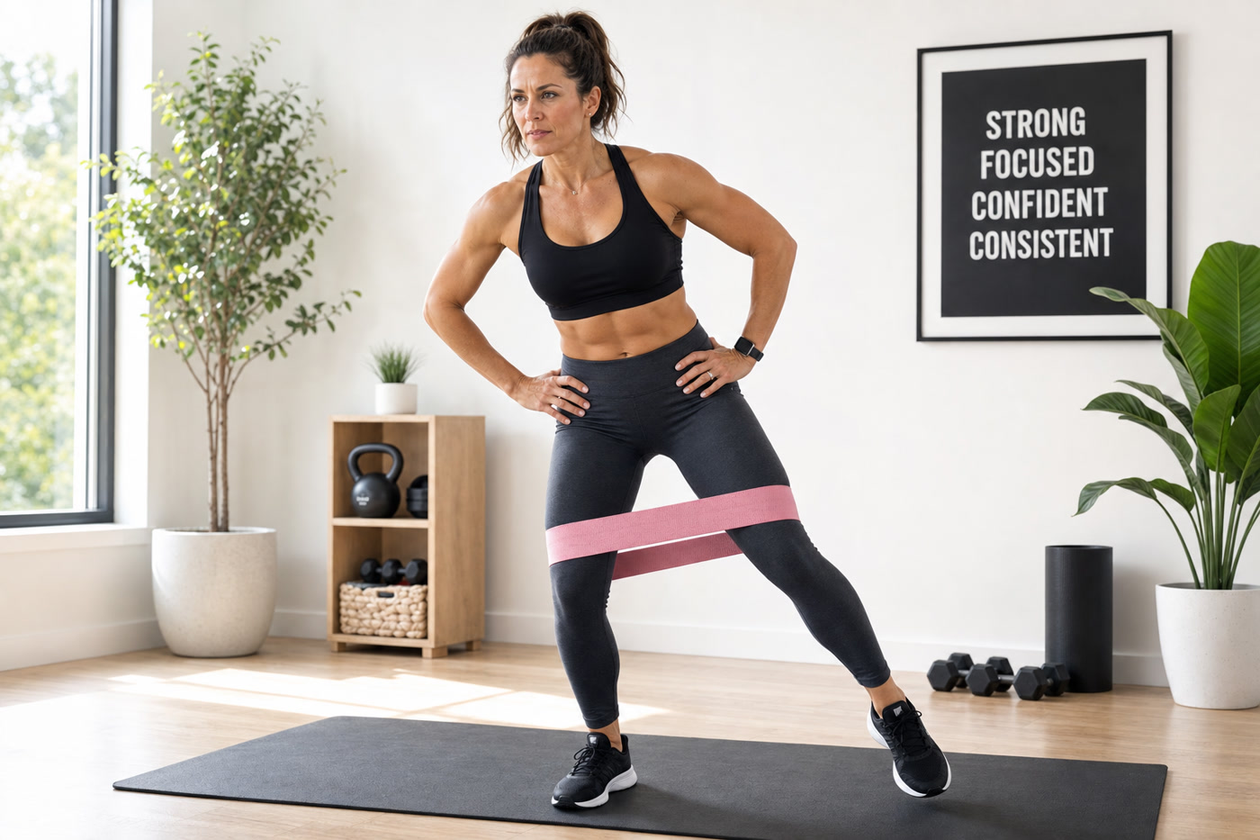
- Gluteus medius activation: Lateral band walks, clamshells, standing hip abduction — building the baseline activation that has likely been absent for years
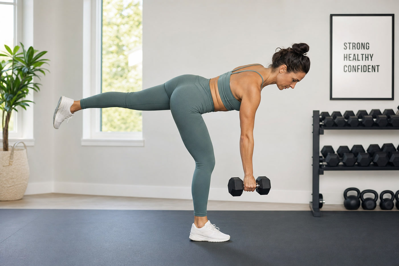
- Gluteus maximus loading: Hip thrusts, single-leg deadlifts, Bulgarian split squats — building the posterior chain strength that controls knee valgus under load
- Hip flexor lengthening: 90/90 stretches, couch stretch, half-kneeling mobilisation — correcting the pelvic position driving APT and glute inhibition
- Graduated knee loading: Controlled squatting with progressive range, step-ups, wall sits — reloading the patellofemoral joint within a pain-free range, progressively expanding that range as hip strength and control improve
- Foot and ankle work: Calf raises, single-leg balance, foot intrinsic exercises — addressing the ground-up component where relevant
- Movement pattern re-education: Retraining squat, lunge, and step mechanics so the knee consistently tracks over the second toe, not caving inward
In-Person at Kitsilano and Downtown Vancouver
TurnFit’s Vancouver studios in Kitsilano and Downtown offer hands-on movement assessment and supervised training. For clients with complex presentations — significant knee pain during most exercise, prior surgeries, or long-standing compensatory patterns — in-person work allows for real-time correction and tactile cueing that accelerates the learning curve.
If you are in Vancouver and want to understand exactly what is driving your knee pain, a TurnFit assessment is the most direct route to that answer.
Train at our Kitsilano or Downtown Vancouver studio. Book a movement assessment with David Turnbull and the TurnFit team — and leave knowing exactly what is causing your pain and what will fix it.
Online Training: Equally Effective, Available Anywhere in Canada
For TurnFit’s clients outside of Vancouver — whether in Calgary, Toronto, or a smaller community where specialist personal training is hard to find — the online training programme delivers the same assessment-driven, hip-dominant protocols.
A 2024 randomised controlled trial published in the International Journal of Exercise Science found that remote and in-person training produced statistically equivalent improvements in strength, body composition, and metabolic health across 12 weeks of identically programmed training. The key ingredient is programming quality and coaching accountability — not physical proximity. (Peña et al., 2024 — Int J Exerc Sci)
TurnFit’s online clients complete their video assessment, receive a written programme with exercise videos and technique cues, and check in regularly with their coach. The same foot-to-pelvis-to-glute assessment framework applies; the delivery simply adapts to the medium.
Not in Vancouver? TurnFit’s custom online training programme delivers evidence-based knee pain solutions from anywhere in Canada.
What to Expect in 6–12 Weeks
One of the most important things we tell new clients at TurnFit is this: your body did not develop this dysfunction overnight, and it will not correct it overnight. But with the right programme, meaningful change happens faster than most people expect.
Weeks 1–2: Activation and Awareness
The first two weeks focus on re-establishing the neuromuscular connection to the glutes. For many women — particularly those who have been desk-bound for years — the gluteus medius and maximus need to be “reminded” how to fire before they can be effectively loaded. Exercises in this phase are low-load, higher-repetition, and focus on activation quality. Pain may not change significantly yet, but the foundation is being built.
Key movements: clamshells, lateral band walks, glute bridges, hip flexor stretching, single-leg balance work.
Weeks 3–4: Loading the Chain
Once activation quality improves, load is introduced progressively. Hip thrusts, step-ups, and split squats enter the programme. Knee loading begins cautiously — partial-range squatting, wall sits — within a pain-free zone. Most clients notice measurable improvements in stair climbing and prolonged walking during this phase.
Key movements: hip thrusts, goblet squats (partial), step-ups, lateral lunges, single-leg deadlifts (light load).
Weeks 5–6: Strength and Movement Pattern Re-education
This is where clinical trials typically measure their outcomes — and where the data shows 50%+ VAS pain reduction in well-designed hip-dominant programmes. Load increases on all hip exercises. Full-range knee exercises are reintroduced where pain permits. Movement patterns are refined: the knee should now be tracking consistently over the second toe rather than collapsing medially.
Key movements: progressive hip thrusts, Bulgarian split squats, Romanian deadlifts, full-range squats, box step-ups with load.
Weeks 7–12: Consolidation and Return to Full Activity
By weeks 7–12, most clients have returned to their previously painful activities with significantly reduced or absent symptoms. Running, group fitness, recreational sport, and workplace stair climbing are re-integrated progressively. Strength continues to build. The programme shifts from rehabilitation to performance — because the goal is not just pain-free daily life; it is a body that can do what you ask of it for decades.
For long-standing or complex presentations, the timeline may extend. But the trajectory is consistent: the right hip-dominant programme, persistently applied, produces cumulative and durable results.
TurnFit also offers dedicated posture and mobility programmes that complement the strength work — particularly for clients whose knee pain has significant APT, hip flexor tightness, or thoracic mobility components.
Frequently Asked Questions
Why do women get more knee pain than men?
Women account for approximately 55% of all PFPS diagnoses and 60% of chondromalacia patella cases in the United States. (Glaviano et al., 2015) Several factors drive this disparity: women tend to have a wider pelvis relative to femur length (a larger Q angle), which creates a stronger inward pull on the kneecap; oestrogen affects ligament laxity across the hormonal cycle; and research shows women demonstrate less gluteus maximus activation during landing tasks compared to men — a neuromuscular difference that allows greater inward knee collapse under load. (Silvers-Granelli, 2021) This biomechanical environment places disproportionate stress on the patellofemoral joint in women, but it is modifiable through targeted training.
What is patellofemoral pain syndrome (PFPS)?
Patellofemoral pain syndrome (PFPS), sometimes called runner’s knee or anterior knee pain, is pain around or behind the kneecap (patella). It is typically aggravated by activities that load the knee in a bent position — squatting, stair climbing, running, cycling, or prolonged sitting. PFPS is not caused by damage to the knee joint itself; it results from the kneecap tracking incorrectly in its groove due to muscle imbalances, particularly weakness in the hip abductors and extensors and poor neuromuscular control of the lower limb. The pain is real; the cause is upstream. The current JOSPT clinical practice guidelines identify hip strengthening as a core evidence-based intervention. (Witvrouw et al., 2019)
Can exercise fix knee pain or will it make it worse?
The right exercise fixes knee pain; rest alone rarely solves it and often allows the underlying muscle weaknesses to worsen. A 2015 multicenter RCT by Ferber et al. involving 199 participants demonstrated significant improvement in both pain and function after just 6 weeks of targeted strengthening (PMID: 25365133). A 2025 meta-analysis confirmed that combined hip and knee strengthening significantly outperforms knee-only exercise (SMD = -1.29, p = 0.0003) (Halabi et al., 2025). The key distinction: exercise that loads a dysfunctional pattern without correcting it can perpetuate symptoms. Exercise that corrects the underlying hip weakness and movement fault directly resolves the pain. An individualised assessment distinguishes between the two.
What is the connection between weak glutes and knee pain?
The gluteus medius is the primary hip abductor — the muscle that prevents the pelvis from dropping and the knee from caving inward (dynamic knee valgus) with every step, squat, or stair. When it is inhibited, the femur rotates inward and adducts, pulling the kneecap out of its tracking groove. A 2025 study found that gluteus medius activation ratio correlated directly with knee function scores in PFPS patients (r = 0.584, p = 0.038): higher glute activation = better knee function. (Ho, Kissman et al., 2025) The gluteus maximus complements this by controlling hip extension and external rotation, reducing dynamic valgus under load and protecting the ACL. (Losciale et al., 2022)
Does my posture at my desk contribute to knee pain?
Yes, significantly. Prolonged sitting tightens the hip flexors and inhibits the glutes through reciprocal inhibition — a pattern that directly undermines the hip control your knee depends on. Research found a moderate positive correlation between anterior pelvic tilt angle and PFPS pain intensity (r = 0.409, p = 0.009): the greater the forward pelvic tilt from sitting, the more intense the knee pain. Workers with knee pain sit an average of 6.72 hours per day. With approximately 3.9 million Canadians diagnosed with osteoarthritis — women at 16.1% versus men at 11.1% — and knee being the most commonly affected joint, the cumulative burden of desk work on knee health over a career is substantial. (Public Health Agency of Canada, 2020) Correcting sitting patterns is helpful; rebuilding hip strength is essential.
What does TurnFit’s assessment involve for someone with knee pain?
TurnFit’s movement assessment evaluates the entire kinetic chain: foot arch posture and pronation, ankle dorsiflexion, tibial rotation, hip strength and activation (gluteus medius and maximus), pelvic position (APT), and lumbar stability. You are observed performing functional movements — single-leg squat, stair descent, step-up — that reproduce the conditions under which your pain occurs. This allows TurnFit to identify which specific links in the chain are failing: is it primarily hip weakness? Foot pronation? APT? All three? From there, a personalised programme is built targeting your exact weak points. Assessments are available in person at Kitsilano and Downtown Vancouver, or online via video for clients across Canada.
How long does it take to reduce knee pain with hip strengthening?
In clinical trials, meaningful pain reduction begins within 4–6 weeks of consistent hip-dominant training. Ferber et al. (2015) demonstrated significant VAS pain reduction over a 6-week protocol (p < 0.001), with the hip group resolving pain one week earlier than the knee-only group. Women in the hip training group achieved an approximately 55% reduction in pain scores at 6 weeks. (Ferber et al., 2015) Most TurnFit clients with patellofemoral knee pain notice functional improvements — easier stair climbing, less post-exercise ache, reduced pain during workouts — within 4–8 weeks. More durable structural changes, and return to full activity including running and strength training, typically occur at 10–12 weeks with a well-adhered programme.
Can I fix knee pain without going to a physio or gym in person?
Yes. A 2024 RCT published in the International Journal of Exercise Science found that remote and in-person training produce statistically equivalent improvements in strength, muscle mass, body composition, and metabolic health over 12 weeks. (Peña et al., 2024) TurnFit’s custom online training programme delivers the same assessment-driven, hip-dominant knee pain protocols used in the Vancouver studios. You do not need specialist equipment: the foundational glute and hip work — clamshells, lateral band walks, glute bridges, hip thrusts — can be done with a resistance band at home. What you do need is a properly individualised programme and consistent coaching accountability.
Is running or squatting safe if I have patellofemoral pain?
Loaded activities do not need to be abandoned — they need to be loaded appropriately while you correct the mechanics causing the pain. Total rest typically worsens PFPS outcomes by allowing hip and quad muscles to weaken further. The clinical approach is to reduce load temporarily (shorten range of motion, reduce volume, use surface modifications), while building hip strength to correct the underlying fault. Once hip control improves and pain levels reduce, full-range squatting and running are progressively reintroduced — usually within 6–12 weeks with proper programming. The goal is not to protect the knee from exercise; it is to make the body strong enough to exercise without producing pain.
What is dynamic knee valgus and why does it matter?
Dynamic knee valgus is the inward collapse of the knee during movement — the “knock-knee” position visible when someone squats or lands from a jump with poor hip control. It matters because it is simultaneously a primary driver of patellofemoral pain and the most significant modifiable risk factor for ACL tears. Women have 3–8 times the ACL injury rate of similarly trained men, and this disparity is substantially explained by neuromuscular patterns — particularly reduced gluteus maximus activation during landing — that allow greater knee valgus under load. (Silvers-Granelli, 2021) Strengthening the gluteus medius and maximus reduces dynamic knee valgus, which simultaneously reduces chronic patellofemoral stress and acute ACL injury risk.
Does foot pronation cause knee pain?
Foot over-pronation can contribute to knee pain through the kinetic chain: when the subtalar joint pronates excessively, the tibia internally rotates, which pulls the patellar tendon medially and disrupts kneecap tracking. Research by Barton et al. (2011) found PFPS patients exhibit earlier peak rearfoot eversion during walking (p = 0.01) — faster foot pronation at heel strike — compared to pain-free controls. (Barton et al., 2011) The JOSPT 2019 PFPS guidelines recommend prefabricated foot orthoses for patients with excessive pronation, specifically combined with exercise therapy. (Witvrouw et al., 2019) Foot pronation is rarely the only cause — it works in combination with hip weakness and ankle mobility limitations — but it must be evaluated as part of any comprehensive knee pain assessment.
I’ve been told to rest my knee — why isn’t it getting better?
Rest reduces the inflammatory signal but does not correct the underlying cause. When you return to activity after resting, you return with the same hip weakness, pelvic tilt, and movement faults — and, often, with weaker muscles than when you started. PFPS has a well-documented tendency toward chronicity under passive management: studies tracking PFPS patients for 5–20 years found that 25–91% continued to report symptoms affecting daily life or physical activity. (Ferber et al., 2018 — J Athl Train) If your knee pain has persisted for more than 6 weeks despite rest, the solution is active rehabilitation targeting the root cause — not more time off. The sooner you begin correcting the hip weakness driving your symptoms, the faster and more durably you will recover.
Ready to Stop Managing Your Knee Pain and Start Fixing It?
Whether you train with us in Kitsilano, Downtown Vancouver, or from your living room in Calgary — TurnFit’s assessment-driven approach to knee pain starts with understanding exactly what is happening in your kinetic chain, and builds from there.
David Turnbull and the TurnFit team have helped hundreds of women go from chronic knee pain and modified workouts to pain-free squats, pain-free stairs, and the confidence to push hard in training without fear. The science supports it. The results confirm it.
References
-
Glaviano NR, Kew M, Hart JM, Saliba S. Demographic and epidemiological trends in patellofemoral pain. Int J Sports Phys Ther. 2015;10(3):281–290.
https://pmc.ncbi.nlm.nih.gov/articles/PMC4458915/ -
Ho KY, Carpio M, Donohue J, Kissman J, Liang JN. Comparison of gluteal muscle central activation in individuals with and without patellofemoral pain. Front Physiol. 2025;16:1535141.
https://www.frontiersin.org/articles/10.3389/fphys.2025.1535141/full -
Ferber R, Bolgla L, Earl-Boehm JE, Emery C, Hamstra-Wright K. Strengthening of the hip and core versus knee muscles for the treatment of patellofemoral pain: a multicenter randomized controlled trial. J Athl Train. 2015;50(4):366–377.
https://pubmed.ncbi.nlm.nih.gov/25365133/ -
Bolgla LA, Earl-Boehm J, Emery C, Hamstra-Wright K, Ferber R. Pain, function, and strength outcomes for males and females with patellofemoral pain who participate in either a hip/core- or knee-based rehabilitation program. Int J Sports Phys Ther. 2016;11(6):926–935.
https://pmc.ncbi.nlm.nih.gov/articles/PMC5095944/ -
Halabi MH, Alturkistani BA, Abuhadi RH, et al. The efficacy of hip and knee muscles strengthening versus knee muscle strengthening alone in managing patellofemoral pain syndrome: a systematic review and meta-analysis. Musculoskelet Care. 2025;23(1):e70059.
https://pubmed.ncbi.nlm.nih.gov/39934098/ -
Silvers-Granelli H. Why female athletes injure their ACL’s more frequently? What can be done to reduce the incidence of ACL injury in female athletes? Int J Sports Phys Ther. 2021;16(4):1208–1213.
https://pmc.ncbi.nlm.nih.gov/articles/PMC8329328/ -
Losciale JM, Bullock GS, Cromwell C, Ledbetter L, Goto S, Pietrosimone L. The influence of gluteal muscle strength deficits on dynamic knee valgus: a systematic scoping review. J Exp Orthop. 2022;9(1):75.
https://pmc.ncbi.nlm.nih.gov/articles/PMC9385941/ -
Barton CJ, Levinger P, Webster KE, Menz HB. Kinematics associated with foot pronation in individuals with patellofemoral pain syndrome: a case-control study. J Foot Ankle Res. 2011;4:18.
https://pmc.ncbi.nlm.nih.gov/articles/PMC3102962/ -
Witvrouw E, Callaghan MJ, Stefanik JJ, et al. Patellofemoral pain: consensus statement from the 4th International Patellofemoral Pain Research Retreat, Manchester, 2017. Part 1: Terminology, definitions, clinical examination, natural history, patellofemoral osteoarthritis and patient-reported outcome measures. J Orthop Sports Phys Ther. 2019;49(9):PG1–PG8.
https://www.jospt.org/doi/10.2519/jospt.2019.0302 -
Peña JC, Martin WF, Cardozo LA, et al. Effects of remote versus in-person training on metabolic profiles and body composition of physically inactive adults: randomized clinical trial. Int J Exerc Sci. 2024;17(4):1016–1025.
https://pmc.ncbi.nlm.nih.gov/articles/PMC11382774/ -
Public Health Agency of Canada. Osteoarthritis in Canada. Government of Canada. Published September 29, 2020.
https://www.canada.ca/en/public-health/services/publications/diseases-conditions/osteoarthritis.html -
Arthritis Research Centre of Canada. British Columbia Osteoarthritis Survey. University of British Columbia.
https://arthritis.rehab.med.ubc.ca/files/2011/08/BCOASurvey.pdf -
Daneshmandi H, Choobineh AA, Ghaem H, Karimi M. Adverse effects of prolonged sitting behavior on the general health of office workers. J Lifestyle Med. 2017;7(2):69–75.
https://pmc.ncbi.nlm.nih.gov/articles/PMC5618737/ -
Ferber R, Bolgla LA, Earl-Boehm JE, Emery CE, Hamstra-Wright K. Treatment success of hip and core or knee strengthening for patellofemoral pain: development of clinical prediction rules. J Athl Train. 2018;53(6):545–552.
https://pmc.ncbi.nlm.nih.gov/articles/PMC6089029/ -
Al-Qattan SI, et al. The Association of Anterior Pelvic Tilt Angle and Clinical Presentations in Patients with Patellofemoral Pain. J Biomed Arthritis Rehabil.
https://jbaar.journals.ekb.eg/article_389599_aa0fd9292c9ea4d4e28112faf08e9d6b.pdf -
Physiopedia. Patellofemoral Pain Syndrome and Hip Strength. Retrieved 2026.
https://www.physio-pedia.com/Patellofemoral_Pain_Syndrome_and_Hip_Strength -
Losciale JM, et al. Journal of Experimental Orthopaedics, 2022 — Scoping review of gluteal muscle strength deficits and dynamic knee valgus.
https://pmc.ncbi.nlm.nih.gov/articles/PMC9385941/ -
Sciascia A, et al. Weaker lower extremity muscle strength predicts traumatic knee injuries in youth female athletes. BMJ Open Sport Exerc Med. 2017.
https://pmc.ncbi.nlm.nih.gov/articles/PMC5731228/ -
JOSPT. Patellofemoral Pain Clinical Practice Guidelines 2019. J Orthop Sports Phys Ther. 2019.
https://www.jospt.org/doi/10.2519/jospt.2019.0302 -
Rathleff MS, et al. Effectiveness of hip muscle strengthening in patellofemoral pain syndrome patients: a systematic review. Braz J Phys Ther. 2015.
https://pmc.ncbi.nlm.nih.gov/articles/PMC4518569/ -
Mündermann A, et al. Effects of lower limb strengthening training on lower limb biomechanical characteristics and knee pain in patients with patellofemoral pain: a systematic review and meta-analysis. Eur J Med Res. 2025.
https://pmc.ncbi.nlm.nih.gov/articles/PMC11807312/ -
Work-Related Musculoskeletal Disorders among Office Workers. Ethiop J Health Sci. 2021.
https://pmc.ncbi.nlm.nih.gov/articles/PMC8047279/ -
JBAAR. Association between patellofemoral pain syndrome and alterations in Q angle and foot pronation. J Phys Med Rehabil Sci.
https://www.jpmrs.org/uploads/332282617229936.pdf -
Movementpi.com. Hip strength predicts non-contact ACL injury. Retrieved 2026.
https://movementpi.com/hip-strength-predicts-non-contact-acl-injury/ -
GLA:D Canada Program for Knee and Hip Osteoarthritis. PLOS ONE. 2023.
https://pmc.ncbi.nlm.nih.gov/articles/PMC10399832/
Ready to Get Started?
Whether you’re in Vancouver or anywhere across Canada, TurnFit builds programs around your life, your goals, and your body. Book your free assessment today — no commitment, just results.
In-person training in Kitsilano & Downtown Vancouver · Custom online training Canada-wide · Corporate wellness programs
See our personal training for women over 40 in Vancouver.
Want to understand the investment before you start? view pricing.
Book your free intro call — no commitment, just results.
Ready to Start Your Fitness Journey?
At TurnFit, we offer in-person personal training at our Kitsilano and Downtown Vancouver locations, online coaching programs with live Zoom calls, and online personal training across Canada. Check out our transparent pricing — no contracts, no hidden fees.
Book a Free 5-Minute Call →